Archive for the HCP Category

Liver Transplantation

Top tips:

  1. Referral to a Transplant Hepatologist should be considered for patients with decompensated cirrhosis, MELD-Na >15, or primary intrahepatic malignancies (hepatocellular cancer).
  2. There are many patient-related factors that can adversely affect patient outcomes. These factors can constitute relative or absolute contraindications to transplantation.
  3. Many transplant programs have made changes to criteria for transplantation in the setting of alcohol-related liver disease (including Alberta). An exception pathway may be available for highly selected individuals who may not have met 6 month abstinence criteria.
  4. Total tumor volume (TTV) and alpha-fetoprotein (AFP) are used to guide liver transplantation eligibility for hepatocellular carcinoma.

General Cirrhosis Admission and Discharge Order Sets

*Add specific panels to general admission orders as appropriate*

For adults with cirrhosis requiring hospital admission
Cirrhosis Adult Admission Orders

For adults with cirrhosis requiring hospital discharge
Cirrhosis Adult Discharge Orders

Thank you to Dr. Sadler for your efforts creating the content on this page!

Liver transplantation (LT) is an accepted therapeutic modality for:

  • End-stage liver disease (acute or chronic); and
  • Selected tumors

Transplantation is indicated for patients with advanced forms of disease in whom no realistic hope for extension of life exists with other forms of medical or surgical intervention. In general, for patients with cirrhosis, one should consider referral for liver transplantation as they develop features of decompensated liver disease such as ascites, bleeding esophageal or gastric varices and hepatic encephalopathy that are not easily controlled with medical/endoscopic therapy.  The MELD-sodium is also a general guide.  Most patients who are considered for transplant have MELD-sodium values >15, but there are certainly exceptions whereby patients with lower MELD scores can be considered. This links to the OPTN (Organ Procurement and Transplantation Network) MELD exception document from 2017.

Indications for Liver Transplantation:

  1. Hepatocellular Liver Disease
    • Nonalcoholic Steatohepatitis
    • Viral Hepatitis
    • Alcohol related Cirrhosis with 6 months abstinence from alcohol.
      • Selected patients may meet criteria for the new exception pathway. Consult Transplant hepatology to discuss these cases.
    • Severe Alcoholic Hepatitis refractory to steroids
      • Selected patients may meet criteria for the new exception pathway. One of the requirements is that there can be no prior history of hospital or ER visits with intoxication or alcohol withdrawal. There are a list of other criteria. Consult Transplant hepatology to discuss these cases.
    • Hepatitis B
    • Cryptogenic Cirrhosis
  2. Cholestatic Liver Disease
    • Primary Biliary Cholangitis, Primary Sclerosing Cholangitis, Secondary Biliary Cirrhosis, Idiopathic or Drug Induced Cholestasis
  3. Inborn Errors of Metabolism
    • Wilson’s Disease, Alpha 1 Antitrypsin Deficiency, Hemochromatosis, Familial Amyloidosis Polyneuropathy, Other
  4. Fulminant Hepatic Failure
  5. Budd-Chiari Syndrome
  6. Drug Toxicity leading to liver failure
  7. Other
  8. Selected intrahepatic malignancies – this refers to tumors originating within the liver, not metastatic disease to the liver. Involve a liver specialist with any suspected diagnosis of Hepatocellular carcinoma (HCC).
    • Listing criteria for Hepatocellular carcinoma
    • Not candidates for transplant
      • TTV >115 cm3 OR AFP >400 ug/mL
      • Metastasis outside the liver
      • HCC vascular invasion on radiological imaging
      • General contraindications for liver transplantation
    • Useful HCC information see HCC page for more HCC details
      • TTV is calculated based on the area of any viable tumor (tumor showing enhancement).
      • Under the guidance of a liver specialist, a patient can be considered for HCC down-staging if TTV ≤250 cm3, regardless of AFP. Following down-staging, a patient can be considered for transplantation if TTV ≤115cm3 AND AFP ≤400 ng/mL for 6 months.
    • TTV calculator

Contraindications for Liver Transplantation

During the evolution of clinical transplantation internationally, and at University of Alberta Hospital, multiple patient-related factors have come to be recognized as adversely affecting patient outcome.   These factors constitute relative or absolute contraindications to transplantation.

The following factors exert a major adverse influence on the outcome of transplantation and would be absolute contraindications in most cases:

Absolute Contraindications

  • Active systemic sepsis
  • Systemic disease such as metastatic cancer, AIDS, mitochondrial disorders
  • pneumonia, unresolved pulmonary infarction or other pulmonary infiltrates
  • Severe non-correctable peripheral or cerebrovascular disease
  • Severe irreversible cardiac disease (unless simultaneous liver and heart transplantation considered)
  • Advanced cardiopulmonary disease
  • Clear history of non-compliance
  • Active alcohol or drug abuse (note: alcohol exception pathway criteria detailed above)
  • Uncontrolled psychiatric disease
  • Severe neurological instability or advanced neurological illness
  • BMI > 45
  • Age > 75 years

Patient materials:

You can direct patients to the following:
Liver transplant

Lab tests

Calculators:

Use these calculators:

Total Tumour Volume Calculation

References:

This section was adapted from content using the following evidence based resources in combination with expert consensus. The presented information is not intended to replace the independent medical or professional judgment of physicians or other health care providers in the context of individual clinical circumstances to determine a patient’s care.

Authors: Dr. Vince Bain, Dr. Matt Sadler, Dr. Rahima Bhanji, Dr. Puneeta Tandon

References:

  1. Adult Liver Transplant Management Guidelines 2016, Version 2.0
  2. Evaluation for Liver Transplantation in Adults: 2013 Practice Guideline by the American Association for the Study of Liver Disease and the American Society of Transplantation. Hepatology 2014 PMID 24716201

ICU Management

Top tips:

When to consult ICU/consider ICU transfer

  1. Hypotension/Shock (SBP < 90 or MAP < 60 with elevated lactate)
  2. Variceal Bleed needing potential airway protection
  3. Significant Acute Kidney Injury (Creatinine > 200 umol/L or worsening trend)
  4. Respiratory failure
  5. Altered mental status (GCS < 10)

General Cirrhosis Admission and Discharge Order Sets

*Add specific panels to general admission orders as appropriate*

For adults with cirrhosis requiring hospital admission
Cirrhosis Adult Admission Orders

For adults with cirrhosis requiring hospital discharge
Cirrhosis Adult Discharge Orders

Check out the bottom of the page for short videos from Dr. Karvellas!

Hypotension/shock:

  • Both circulatory and cardiac abnormalities may develop in patients with cirrhosis
  • In cirrhosis/acute on chronic liver failure (ACLF) patients presenting with shock, the approach should be guided by a systematic assessment of volume status and correction of hypotension in an effort to restore adequate end organ perfusion.
  • Arterial catheters are recommended to guide resuscitative efforts. Central venous access is also recommended to aid the assessment of hemodynamic status (e.g. measurement of central venous pressure (CVP)) and as a route for vasoactive medications.
  • Assessment of volume status in this patient population can be particularly challenging due to ascites and edema. Dynamic measures of volume and circulatory function such as echocardiography, changes in CVP in response to fluid challenge are preferable
  • In ACLF/cirrhosis patients with additional organ failures aim for a MAP > 65 mmHg
  • Volume expansion with ringer’s lactate, plasmalyte or concentrated albumin (25% 100 cc prn) is appropriate with fluid choice guided by the patient’s clinical status
  • With upper GI bleeding, there is evidence to support a restrictive transfusion threshold of 70 g/L. This threshold is liberalized if bleeding is massive or if there is a history of ischemic heart disease etc.
  • Norepinephrine is the recommended first-line agent as it is associated with fewer adverse events. Vasopressin may be used as a second-line agent.
  • In patients where adrenal insufficiency is suspected, it is our practice to empirically administer hydrocortisone 200 mg IV in 4 divided doses. Continue until resolution of shock.

Respiratory failure:

  • Pulmonary complications in cirrhosis can either be directly related to cirrhosis (portopulmonary hypertension, hepatopulmonary syndrome, hepatohydrothorax) or unrelated (e.g. pneumonia)
  • Tense ascites and hepatic hydrothorax may complicate mechanical ventilation.
  • Use typical lung protective ventilation strategies with low tidal volume ventilation and use of positive end expiratory pressure (PEEP) to maintain oxygenation.
  • In patients with unexplained hypoxia, consider diagnostic studies to evaluate for hepatopulmonary syndrome and portopulmonary hypertension (e.g. contrast echocardiography).

Neurological Dysfunction

  • See Hepatic Encephalopathy section for more on the topic
  • Treat possible precipitating factors for hepatic encephalopathy (HE) and evaluate the response to ammonia lowering therapies (lactulatose, PEG, rifaximin).
  • Consider CT head in patients do not respond to standard treatments or where HE onset is abrupt or severe
  • Consider electroencephalogram (EEG) to exclude other causes of altered mental status in patients who fail to respond to standard therapy
  • Consider intubation for airway protection in patients with a GCS of ≤ 8
  • In patients who require intubation/mechanical ventilation for respiratory failure, the use of short acting agents such as fentanyl (25-200 ug/hour) or propofol (50-150 mcg/kg/min) should be considered. Avoid benzodiazepines as these agents precipitate more pronounced neurocognitive impairment

 Renal Dysfunction

  • See Renal Dysfunction section for more on the topic
  • Hepatorenal syndrome (HRS) and its physiology of renal vasoconstriction and splanchnic vasodilatation are one of several causes of Acute Kidney injury (AKI) in cirrhosis. Other causes of AKI include hypovolemia/prerenal azotemia, intrinsic renal/parenchymal disorders (acute tubular necrosis/ATN, nephrotoxicity, interstitial nephritis, glomerulonephritis/ nephropathy), and obstructive nephropathy. Consider urinalysis, renal ultrasound, blood pressure evaluation and medication history to rule out other causes of AKI.
  • Albumin (1g/kg (20-25%) on day one, then 20-60 g/day thereafter) along with vasoconstrictor therapies have been relatively well studied in the treatment of HRS, in particular, vasopressin Consider Midodrine (maximum dose 15 mg TID) and octreotide (maximum dose 200 ug sc TID) if on the ward. If no effect, then a therapeutic trial of IV norepinephrine to maintain a MAP > 75 mm Hg in the ICU can be considered.
  • Intermittent or continuous renal replacement therapy (RRT) may be used in HRS as a bridge to LT. The use of RRT in patients with HRS is likely only appropriate in the setting of a patient who is listed for LT or has another indication for RRT (i.e. uremia, acidosis, hyperkalemia)

Hemostatic Disorders:

  • Patients with cirrhosis are in a fragile continuum between ineffective hemostasis and inappropriate thrombosis due to decreased production of procoagulant and anticoagulant factors
  • INR (international normalized ratio) does not account for deficiencies of the anti-coagulation system (especially low protein C), which may result in a hypercoagulable state not reflected in prolongation of the INR.
  • Consider maintaining platelets > 50 and a fibrinogen > 1.5 g/L as targets in cirrhosis patients requiring procedures or with active bleeding
  • If available, consider viscoelastic tests of whole blood (thromboelastography (TEG), thromboelastometry (ROTEM)).

Variceal Bleeding

  • See Varices section for more on the topic
  • Consider early intubation in cirrhosis/ACLF patients with concomitant HE, respiratory distress or large volume hematemesis (> 500 ml blood) prior to performing endoscopy. Consider octreotide infusion to improve portal inflow with early endoscopy.
  • In the event of failed endoscopy, a balloon tamponade device, such as the Sengstaken-Blakemore or Minnesota tube may be used as a bridge to more definitive therapy including repeat endoscopy or transjugular intrahepatic portosystemic shunting
  • Administer prophylactic antibiotics (i.e. ceftriaxone 1g IV qd) given potential reduction in rates of rebleeding and bacterial infections

Sepsis

  • See Spontaneous Bacterial Peritonitis and Pleuritis section for more on the topic
  • Early antibiotics in cirrhosis/ACLF patients with septic shock improves mortality
  • Increasing numbers of ACLF patients are presenting with septic shock bacteremia from gram negatives (50-60%) and multidrug resistant pathogens
  • If severe sepsis is suspected, a thorough evaluation should promptly be followed by antibiotic administration, since each hour delay impairs outcome
  • In patients with clinical improvement within 48-72 hours and a known pathogen, immediate tailoring of antibiotics is recommended. In patients without clinical improvement empiric antifungal therapy and CT scan is warranted.

Acute-on-chronic liver failure (ACLF) prognostication

Organ SystemScore = 1Score = 2Score = 3
Liver (mg/dl)Bilirubin < 66 ≤ Bilirubin ≤ 12Bilirubin >12
Kidney (mg/dl)Creatinine <2Creatinine ≥2 <3.5Creatinine ≥3.5 or
renal replacement
Brain
(West-Haven)
Grade 0Grade 1-2Grade 3-4
CoagulationINR < 2.02.0 ≤ INR < 2.5INR ≥ 2.5
CirculationMAP ≥70 mm/HgMAP ≥70 mm/HgVasopressors
Respiratory: PaO₂/FiO₂
or SpO₂/FiO₂
>300
>357
≤300 - > 200
>214- ≤357
≤200
≤214

A CLIF-ACLF score of > 70 is associated with 90% mortality at Day 28. While predicting futility in ACLF is challenging, decisions regarding ICU admission should be based on factors including:

  • Goals of Care
  • Candidacy for liver transplant
  • Age
  • CLIF-C ACLF score
  • Other comorbidities/sarcopenia

Introducing Dr. Karvellas

Video 1 – Common situations where you should consider calling ICU to assist with the management of a hospitalized patient with cirrhosis.

Video 2 – Why it is essential that you have a goals of care conversation with all of your patients and practical tips about how and when to use the acute-on-chronic liver failure calculators.

Patient materials:

You can direct patients to the following:
When to go to emergency 

Downloadable content:

You can download these to print or view offline:
AASLD Guidelines for HE

EASL Guidelines for decompensated cirrhosis

References:

This section was adapted from content using the following evidence based resources in combination with expert consensus. The presented information is not intended to replace the independent medical or professional judgment of physicians or other health care providers in the context of individual clinical circumstances to determine a patient’s care.

Authors: Dr. Dean Karvellas, Dr. Puneeta Tandon

References:

  1. Cecconi M et al. Consensus on circulatory shock and hemodynamic monitoring. Task force of the European Society of Intensive Care Medicine. Intensive Care Med 2014;40:1795-1815. PMID 25392034
  2. De Backer D et al. Comparison of dopamine and norepinephrine in the treatment of shock. N Engl J Med 2010;362:779-789.PMID 20200382
  3. Fernandez J et al. Adrenal insufficiency in patients with cirrhosis and septic shock: Effect of treatment with hydrocortisone on survival. Hepatology 2006;44:1288-1295. PMID 17058239
  4. Ventilation with lower tidal volumes as compared with traditional tidal volumes for acute lung injury and the acute respiratory distress syndrome. The Acute Respiratory Distress Syndrome Network. N Engl J Med 2000;342:1301-1308. PMID 10793162
  5. Nanchal R et al. Guidelines for the Management of Adult Acute and Acute-on-Chronic Liver Failure in the ICU: Cardiovascular, Endocrine, Hematologic, Pulmonary, and Renal Considerations. Critical Care Medicine 2020; 48(3):173-191. PMID 32058387.
  6. Rahimi RS et al. Lactulose vs polyethylene glycol 3350–electrolyte solution for treatment of overt hepatic encephalopathy: the HELP randomized clinical trial. JAMA Intern Med 2014;174:1727-1733. PMID 25243839
  7. Nassar Junior AP, Farias AQ, LA DA, Carrilho FJ, Malbouisson LM. Terlipressin versus norepinephrine in the treatment of hepatorenal syndrome: a systematic review and meta-analysis. PloS one 2014;9:e107466. PMID 25203311
  8. Villanueva C et al. Transfusion strategies for acute upper gastrointestinal bleeding. N Engl J Med 2013;368:11-21. PMID 23281973
  9. Arabi YM et al. Antimicrobial therapeutic determinants of outcomes from septic shock among patients with cirrhosis. Hepatology 2012;56:2305-2315. PMID 22753144
  10. Jalan R et al. CANONIC study investigators of the EASL-CLIF Consortium. Development and validation of a prognostic score to predict mortality in patients with acute-on-chronic liver failure. J Hepatol. 2014;61(5):1038-47. PMID 24950482

Portal Vein Thrombosis

Top tips:

  1. Available data on this topic is predominantly non-randomized and observational. Guidance statements are consistent with expert documents from AASLD (PMID 33219529, Baveno VII (PMID 35120736) and PMID 33930474). Consult specialist on-call for most cases of PVT in cirrhosis.
  2. The main potentially lethal consequences of PVT in cirrhosis are variceal hemorrhage and intestinal ischemia/necrosis
  3. Doppler ultrasound is the first-line evaluation for the diagnosis of portal vein thrombosis (PVT). Perform an Ultrasound doppler to evaluate for PVT in all patients with (i) a new diagnosis of cirrhosis, (ii) onset of portal hypertension, (iii) hepatic decompensation.
  4. CT or MRI should be used to confirm the diagnosis, to exclude tumor thrombus and assess extension into the mesenteric veins
  5. In patients with chronic PVT who meet indication for anticoagulation, assess for high-risk varices with endoscopy. If high risk varices are present, non-selective beta-blockers are suggested for the primary prevention of variceal bleeding.
  6. In cirrhosis, it can be challenging to determine the chronicity of PVT because portal cavernoma is less frequent.
  7. The goal of PVT therapy in cirrhosis is to avoid progression of the clot that predisposes to worsening portal hypertension, impacts the ability to perform a liver transplant, or liver transplant outcomes.
  8. Variceal prophylaxis should be carried out as per AASLD and Baveno variceal guidelines. Link to varices page
  9. See algorithm below for indications for anticoagulation.
  10. HCC related tumor thrombus does not benefit from anticoagulation
  11. Continue anticoagulation until recanalization, and for at least 6 months. Consider longer term anticoagulation even after recanalization in all patients, especially those with SMV involvement, a history of intestinal ischemia or thrombophilic state or if the patient is a liver transplant candidate.
  12. If anticoagulation is stopped, check PV patency within 3 months after stopping, as re-thrombosis rates are high – up to 38% by 4 months.

General Cirrhosis Admission and Discharge Order Sets

*Add specific panels to general admission orders as appropriate*

For adults with cirrhosis requiring hospital admission
Cirrhosis Adult Admission Orders

For adults with cirrhosis requiring hospital discharge
Cirrhosis Adult Discharge Orders

Thank you to Dr. Garcia-Tsao for your efforts creating the content on this page!

Patient materials:

You can direct patients to the following:
Portal hypertension

Downloadable content:

You can download these to print or view offline:
EASL Guidelines for decompensated cirrhosis

References:

This section was adapted from content using the following evidence based resources in combination with expert consensus. The presented information is not intended to replace the independent medical or professional judgment of physicians or other health care providers in the context of individual clinical circumstances to determine a patient’s care.

Authors:  Dr. Vladimir Marquez, Dr. Guadalupe Garcia-Tsao, Dr. Wayne Bai, Dr. Puneeta Tandon

References:

  1. Simonetto DA, Singal AK, Garcia-Tsao G, Caldwell SH, Ahn J, Kamath PS. ACG Clinical Guideline: Disorders of the Hepatic and Mesenteric Circulation. Am J Gastroenterol 2020 Jan;115(1):18-40. PMID 31895720
  2. Francoz C, Valla D, Durand F. Portal vein thrombosis, cirrhosis, and liver transplantation. J Hepatol 2012;57:203–12. PMID 22446690
  3. Nery F, Chevret S, Condat B, et al. Causes and consequences of portal vein thrombosis in 1,243 patients with cirrhosis: Results of a longitudinal study. Hepatology 2015;61:660–7.PMID 25284616
  4. Yerdel MA, Gunson B, Mirza D, et al. Portal vein thrombosis in adults undergoing liver transplantation: Risk factors, screening, management, and outcome. Transplantation 2000;69:1873–81.PMID 10830225
  5. Kreuziger LB, Ageno W, Lee A. Management of incidental splanchnic vein thrombosis in cancer patients. Hematology Am Soc Hematol Educ Program 2014;2014:318–320. doi: 10.1182/asheducation-2014.1.318.PMID 25696872

Last reviewed November 3, 2022

Alcohol Associated Hepatitis (Alcoholic hepatitis)

Top tips:

  1. Rule out infection and additional causes of jaundice in all patients with suspected alcohol associated hepatitis
  2. Severe alcohol associated hepatitis is most easily diagnosed by a MELD >20 (this is the original MELD calculation, not the MELD-Na)
  3. Need to consider the management of alcohol withdrawal, nutritional therapy as well as the treatment of alcohol use disorder alongside the treatment of alcohol associated hepatitis
  4. Use the Lille model at 7 days to determine if the patient is a non-responder to steroid therapy (Lille ≥ 0.45)
  5. Using very strict criteria, the Alberta transplant program is now evaluating some patients with alcohol associated hepatitis for liver transplantation.

Order panel for Alcohol associated hepatitis:

For adults admitted with Alcohol associated hepatitis.
Alcohol Associated Hepatitis Order Panel

General Cirrhosis Admission and Discharge Order Sets

*Add specific panels to general admission orders as appropriate*

For adults with cirrhosis requiring hospital admission
Cirrhosis Adult Admission Orders

For adults with cirrhosis requiring hospital discharge
Cirrhosis Adult Discharge Orders


Thank you to Dr. Borman for your efforts creating the content on this page!

Diagnosis

General Management

Specific Management

Patient materials:

You can direct patients to the following:
Alcohol

211.ca

AHS Help in Tough Times

Alberta detox and addiction treatment centre options

Calculators:

Use these calculators to help with the diagnosis:

lillescoreINR

www.alchepscores.com

original MELD score

Downloadable content:

You can download these to print or view offline:
AASLD guidance statement 2019

References:

This section was adapted from content using the following evidence based resources in combination with expert consensus. The presented information is not intended to replace the independent medical or professional judgment of physicians or other health care providers in the context of individual clinical circumstances to determine a patient’s care.

Authors: Dr. Meredith Borman, Dr. Monty Ghosh, Dr. Puneeta Tandon

References:

  1. Crabb DW et al. Diagnosis and Treatment of Alcohol-Related Liver Diseases: 2019 Practice Guidance from the American Association for the Study of Liver Diseases. Hepatology 2019 July 17 epub ahead of print, PMID 31314133
  2. Singal AK et al. ACG Clinical Guideline: Alcoholic Liver Disease. Am J Gastroenterol 2018 Feb;113(2):175-194 PMID 29336434
  3. Nguyen-Khac N et al. Glucocorticoids plus N-acetylcysteine in severe alcoholic hepatitis. N Engl J Med 2011 Nov; 365(19):1781-9 PMID 22070475

Hepatocellular Carcinoma

Top tips:

  1. HCC is the sixth most common cancer, the third leading cause of cancer-related deaths, and the second most lethal cancer. It represents 90% of primary liver cancers and occurs in the presence of underlying cirrhosis most of the time.
  2. Patients with chronic HBV infection and patients with NAFLD/MAFLD can develop HCC in the absence of cirrhosis
  3. Patients with cirrhosis (Child Pugh A or B or those patients with Child Pugh C awaiting liver transplantation) and patients without cirrhosis with chronic HBV infection at increased risk of HCC should undergo HCC surveillance with ultrasound and AFP every six months.
  4. The diagnosis of HCC in patients with cirrhosis is based on dynamic imaging (e.g., CT, MRI or CEUS) according to well-defined criteria. Liver biopsy is needed to confirm the diagnosis in cases with atypical imaging features and in all patients without underlying cirrhosis.
  5. Staging and treatment allocation are based on the Barcelona Clinic Liver Cancer (BCLC) staging system, which takes into account the tumor burden, the underlying liver function, and the patient’s performance status.
  6. All patients with HCC must be reviewed at a Multi-Disciplinary Team (MDT) meeting before selecting therapy. This approach has increased survival rates in other series. Within Alberta, this occurs in Edmonton and Calgary.

General Cirrhosis Admission and Discharge Order Sets

*Add specific panels to general admission orders as appropriate*

For adults with cirrhosis requiring hospital admission
Cirrhosis Adult Admission Orders

For adults with cirrhosis requiring hospital discharge
Cirrhosis Adult Discharge Orders

Dr. Burak cartoon
Dr. fung cartoon
Untitled design (5)

Thank you Dr. Burak, Dr. Fung, and Dr. Moctezuma Velazquez for your efforts in creating content for this page. Check out the bottom of the page for short videos from Dr. Fung and Dr. Burak!

Risk factors and prevention

  • The most important risk factor for HCC is liver cirrhosis. Hence, to prevent progression to cirrhosis, it is very important to detect chronic liver diseases in the general population early on in their natural history. Other risk factors for HCC are age, male sex, diabetes, obesity, alcohol, tobacco, and aflatoxin B1 exposure.
  • Primary prevention should aim to prevent chronic liver disease by promoting a healthy diet, exercise, responsible drinking, and HBV vaccination.
  • Secondary prevention should target the treatment of the etiology of the underlying liver disease, and other risk factors associated with HCC, in order to prevent disease progression and development of HCC. Coffee drinking may protect against HCC.

Note: Tertiary prevention would aim to reduce the risk of recurrence after curative treatments, but so far, there are no neoadjuvant or adjuvant therapies approved for this purpose. 

Surveillance

Who should be screened:

  • Patients with cirrhosis (regardless of the etiology) who are Child Pugh A or B.
  • Given limited treatment options, surveillance should only be done in Child Pugh C patients if they are eligible for liver transplantation
  • Patients with HBV infection without cirrhosis belonging to one of the following groups:
    • PAGE-B score ≥10
    • Asian females >50 years
    • Asian males >40 years
    • Family history of HCC
    • Patients of African descent >20 years

Note: Patients with HCV-associated cirrhosis should continue to undergo screening even after SVR.

How to screen:

  • Ultrasound surveillance with serum alpha-fetoprotein (AFP) every six months. Overall sensitivity of the ultrasound is 84%, but only 45% for early stages; the addition of AFP increases sensitivity to 63% in early stages.
  • Do not use AFP alone for surveillance
  • For ultrasound/AFP to be cost-effective, the adherence needs to be ≥34%, and the annual incidence of HCC should be ≥1.5% and ≥0.3% in patients with and without cirrhosis, respectively. Automated radiology recall programs are preferred to increase compliance – for Alberta, please see details for these programs in the grey bar.
  • If there is a mass/nodule on the ultrasound > 1cm, patients need to undergo a dynamic imaging study (see diagnosis below).
  • If there is a lesion < 1 cm, the ultrasound should be repeated at shorter intervals (e.g. every 3-4 months). If the lesion remains stable after 12 months of follow-up, the patient can return to every 6 months for surveillance. On the contrary, if the lesion grows or changes in pattern, the patient will need to undergo a dynamic imaging study (see diagnosis below).

NOTE: Ultrasound may perform sub-optimally in patients with obesity, steatosis, and/or multinodular livers. The US LI-RADS visualization score (i.e. A for minimal limitations, B for moderate limitations, and C for severe limitations) can help identify the need for an alternative screening strategy in a given patient (e.g. cross-sectional imaging).

Diagnosis

HCC
  • In patients with underlying cirrhosis, imaging criteria defined by the European Association for the Study of the Liver (EASL) and Liver Imaging Reporting and Data System (LI-RADS) enable HCC diagnosis (without the need for targeted biopsy).
  • A lesion >1cm showing arterial phase hyperenhancement and non-peripheral venous washout can be diagnosed as HCC; LI-RADS criteria also take into account threshold growth (>50% diameter increase in <6 months).
  • In patients with cirrhosis and non-conclusive imaging findings, a multidisciplinary discussion is suggested to decide the best strategy to follow: targeted biopsy, close follow-up, or repeated imaging with an alternative method.
  • Liver biopsy is mandatory in non-cirrhotic patients given the lower pre-test probability of HCC.
  • If a biopsy is done, specific stains may help with the diagnosis of HCC.
  • Isolated alpha-fetoprotein (AFP) elevation is not required for the diagnosis of HCC (40% of HCC do not make AFP)
  • AFP should not be used on its own to diagnose HCC (mixed tumors can also produce AFP).
  • AFP should be ordered if HCC is suspected as it has prognostic properties and is used to define liver transplant and systemic therapy (e.g. ramucirumab) candidacy.


Total Tumour Volume Calculation

Radiology findings

Staging and Treatment – the Alberta HCC Algorithm

    • Once the diagnosis of HCC is confirmed, a CT of the chest should be obtained for starting.
    • If there is any suspicion of bone metastasis (e.g. bone pain), a bone scan should also be obtained.
    • Staging and treatment allocation are based on the BCLC staging system, which takes into account the tumor burden (e.g. number, size, vascular invasion, extrahepatic spread), the underlying liver function, and the patient´s performance status (PS).
    • The BCLC comprises 5 stages:
Very early stage (0):

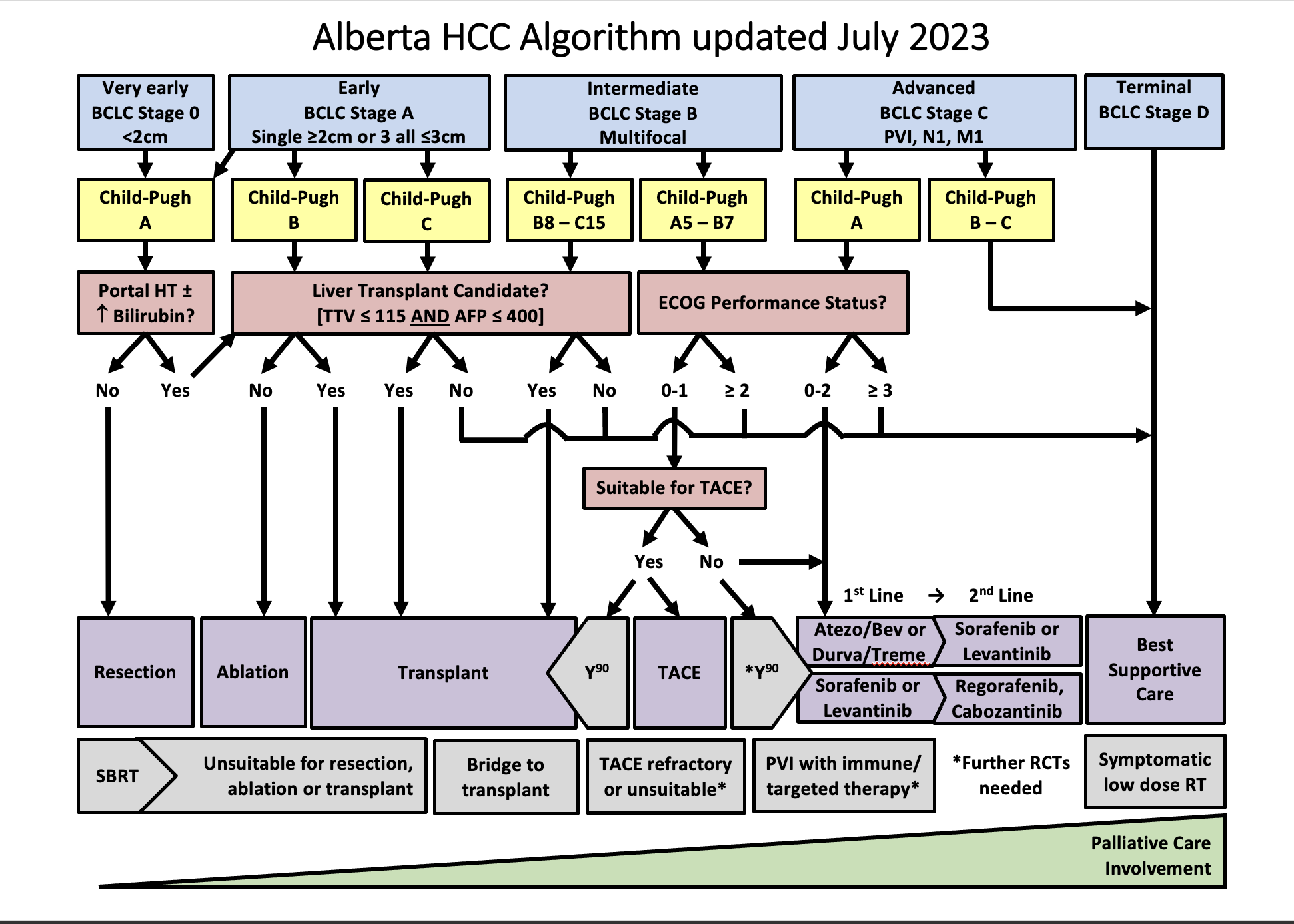
Defined as a single lesion <2 cm in a patient with preserved liver function, without vascular invasion, extrahepatic spread, or cancer related symptoms (PS=0). The most recommended treatment for these patients is ablation, but resection is another option. Ablation and resection offer similar survival rates. These patients are not granted exception points for liver transplantation. Other treatment options, when ablation and resection are not possible, are TACE, TARE, and SBRT.

Early stage (A):

Defined as a single lesion >2cm, or a multifocal HCC with up to 3 nodules, each < 3 cm. In addition, patients need to have preserved liver function and should be free of vascular invasion, extrahepatic spread and/or cancer related symptoms (PS=0). The preferred treatment for single lesions, if possible, is resection. Liver transplant is the treatment of choice for patients with multifocal HCC or with solitary HCC < 5 cm not amenable to resection; patients that are not eligible for liver transplant should undergo ablation. If a patient with HCC is expected to be on the waiting list for > 6 months, the HCC should receive treatment as a bridging strategy to liver transplant to prevent HCC progression beyond transplant criteria. There is evidence to support the use of ablation, TACE, or TARE, or SBRT as a bridge to liver transplant. Single lesions < 8 cm can also be treated with TARE. Another alternative, when the previous strategies are not feasible, is the use of SBRT.

Intermediate stage (B):

Refers to patients with preserved liver function, without vascular invasion, extrahepatic spread, or cancer related symptoms (PS=0), but with a tumor burden beyond that of stage A. This is a very heterogeneous stage, and can be subdivided in 3 groups:

1.Patients with a tumor burden beyond transplant criteria, but with a  well defined disease that is within criteria to allow downstaging strategies. These criteria are defined by each center or transplant program. In Alberta, a patient can be considered for down-staging IF the TTV is ≤ 250 cm³, regardless of AFP. Following downstaging, a patient can be considered for transplantation if TTV ≤ 115 cm³ and AFP ≤400 ug/mL for 6 months. Downstaging can be achieved with ablation, TACE, or TARE.

2. Patients with a well defined tumor burden that is amenable to TACE, and not considered for LT. Another treatment option is TARE, which is better suited for single lesions. SBRT can also be considered.

3.Patients with diffuse, infiltrative, and/or extensive tumor burden not suitable for TACE. These patients are better served with systemic therapy.

Advanced stage (C):

Defined by vascular invasion and/or extrahepatic spread and/or minor cancer related symptoms (PS=1-2). These patients should be treated with systemic therapy.

Terminal stage (D):

Comprises patients with compromised liver function not being considered for LT, and/or patients with major cancer-related symptoms(PS>2). These patients should receive best supportive care.

Reproduced with permission from Dr. K. Burak. Source: HepAPPtology

Child Pugh Classification
Factor 1 point 2 points 3 points
Total Bilirubin (μmol/L) <34 34-50 >50
Serum Albumin (g/L) >35 28-35 <28
PT INR <1.7 1.71-2.30 >2.30
Ascites None Controlled, on diuretics Refractory
Hepatic Encephalopathy None Controlled, on medical therapy Refractory

ECOG Performance Status (PS)
PS Description
0 Fully active and able to carry on without restrictions
1 Unable to carry out physically strenuous activities but ambulatory and able to complete work of a light or sedentary nature
2 Ambulatory and capable of all self-care but unable to complete work activities. Up and about more than 50% of waking hours
3 Capable of only limited self-care and/or confined to a bed or chair for more than 50% of waking hours.
4 Completely disabled. Unable to carry out any self-care. Totally confined to a bed or chair.

Surgical Resection
  • Must have preserved liver function (Child Pugh A) with no significant portal hypertension (no varices, hepatic venous pressure gradient or HVPG < 10 mmHg, platelets >100 or the combination of LSM-TE < 15 kPa and platelets > 150) and a normal bilirubin to avoid post-op liver failure
  • Recurrence occurs in 70% at 5 years
  • RCTs vs RFA show similar survival but fewer recurrences after surgery in the early stage
  • Complications include post-op infections and liver failure (2% mortality)
  • Laparoscopic resection has a lower rate of postoperative complications and can be considered in selected cases even in patients with clinically significant portal hypertension
Ablation (Radiofrequency ablation [RFA], Microwave ablation [MWA], or Percutaneous ethanol injection [PEI])
  • Lesions should be <3cm and there should be < 3 lesions
  • Can be performed in Child Pugh A or B (those who are not candidates for surgery)
  • Meta-analyses of RCTs show that RFA is better than PEI for local control & disease free survival if tumour >2cm but PEI is preferred if concerns about thermal injury to adjacent structures or proximity to blood vessels where heat-sink can be an issue with RFA
  • Compared to RFA, MWA achieves more tumor necrosis and is less prone to heat sink effect
  • Recurrence also 70% at 5yrs (>2.5 cm predicts recurrence)
  • Complications in 2-5%: bleeding, thermal injury, abscess, tumour seeding (1%)
Liver Transplantation
  • LT is the only therapy that treats both the HCC and underlying cirrhosis, so it can be done in Child Pugh C
  • Milan criteria [single <5cm, 3 HCC <3cm] predicts low risk for recurrence (<15%)
  • Patient require long-term immunosuppression, which has many complications, and organ donor shortage lead to many patients progressing beyond accepted criteria while waiting for LT
Transarterial chemoembolization (TACE)
  • Takes advantage of the rich arterial blood supply of tumours to provide chemotherapy (usually doxorubicin) with embolic particles (induces tumour ischemia)
  • Need to have good liver function (CPA or CPB7) to prevent liver failure from ischemia, and must avoid if there is a main portal vein (PV) thrombosis (chance of liver failure)
  • Caution if bilirubin >34 umol/L or ascites especially if not a candidate for transplantation
  • Consideration should be given to systemic therapy for tumor burden beyond up to seven criteria, because these tumors may be associated with worse outcomes after TACE.
  • Use of drug eluting beads (DEB-TACE) provides a more standardized technique and is better tolerated (but more expensive), but offers no survival advantage
  • Complications include post embolization syndrome (fever, nausea, vomiting) in approximately 25%, ischemic complications and rarely liver failure (2-3% mortality)
Transarterial radioembolization (TARE)
  • Like TACE it should be reserved for CPA or CPB7
  • Yttrium (Y90) microspheres provide internal radiation to the tumour
  • Particles are smaller and less embolic and can be used if PV thrombosis
  • Cohort studies and small RCTs show longer time to progression when compared to TACE, but with no survival advantage
  • May be better than TACE for large tumours and for bridging patients to LT
  • It is more expensive than TACE and requires two angiograms (first to calculate shunt fraction to lungs and to embolize collaterals) but it can be done as an outpatient as post embolization syndrome is uncommon.
  • Better results with personalized dosimetry and when targeting single lesions
  • Complications include radiation damage to liver, lungs or GI tract. TARE Referral form
Stereotactic body radiation therapy (SBRT)
  • Because there is a lack of robust evidence, SBRT is usually reserved for patients who aren’t candidates for other locoregional therapies
  • Provides confocal external radiation in 5 fractions
  • Cohort studies show good local control
  • Complications include radiation damage to liver
  • Other SBRT considerations from the Cancer Institutes in Edmonton and Calgary:
    • Good performance status (ECOG 0 -2)
    • Life expectancy >6 months
    • Limited number of metastases (<3-5)
    • Limited prior overlap RT
    • Patient able to tolerate RT position
    • Limited tumor size (< 6 cm)
    • Controlled or absent extra hepatic disease
    • At least 700 cc of uninvolved liver volume
    • Adequate liver function (Child Pugh A)
    • Adequate hematologic function (Absolute neutrophil count (ANC) ≥ 1,500 cells/mm3, Platelets ≥ 70,000 cells/mm3, Hemoglobin ≥ 8.0 g/dl)
  • Additional contraindications to radiotherapy
  1. Tumor location close to radiosensitive structures like the stomach, small bowel, large bowel, kidney
  1. Ascites - poor reproducibility and represents poor liver reserve

Systemic therapy

Atezolizumab and Bevacizumab
  • Combination of immunotherapy and targeted therapy
  • Atezolizumab (monoclonal antibody checkpoint inhibitor that blocks PD-L1) and bevacizumab (monoclonal antibody that blocks VEGF) showed statistically improved overall survival
  • When compared to sorafenib [19.2 vs 13.4 months, HR 0.66 (95% CI: 0.52, 0.85)], and also a longer time to deterioration of quality of life (11.2 vs 3.6 months).
  • First line systemic therapy of choice
  • Patients need to have a recent upper endoscopy and if varices are present, adequate prophylaxis should be in place before first starting treatment.
  • Compared to sorafenib, patients have more hypertension and proteinuria, but less diarrhea and hand-foot skin reaction.
  • Contraindicated in liver transplant recipients
  • Caution in patients with autoimmune conditions
Sorafenib
  • Targeted oral therapy with anti-VEGF (vascular endothelial growth factor), PDGF (platelet derived growth factor), RAF kinase activity
  • Can be used as a first line systemic therapy when there is a contraindication for atezolizumab-bevacizumab
  • RCTs have shown a survival benefit over placebo in patients with preserved liver function (Child Pugh A)
  • Side-effects include nausea, diarrhea, hand & foot skin reactions and hypertension
  • There is real-world evidence to support its use in Child-Pugh B patients
  • Can be used after liver transplant
Lenvatinib
  • Targeted oral therapy that blocks VEGF receptors, FGF (fibroblast growth factor) receptors, PDGF receptor, RET, and KIT
  • Found to be non-inferior to sorafenib in large open-label phase 3 study (median
  • Survival 14 months for levatinib vs 12 months for sorafenib) with higher tumour response rates. Trial excluded patients with main portal vein invasion and/or bulky tumors (>50% of liver parenchyma involved).
  • Can be used as a first line systemic therapy when there is a contraindication for atezolizumab-bevacizumab
  • Side-effect profile similar to sorafenib
Regorafenib
  • Targeted oral therapy against FGFR, VEGFR, RAF, TIE2, PDGFR
  • Approved as a second line therapy after progression with sorafenib, it improved survival compared to placebo [10.6 vs 7.8 months, HR 0.63 (0.50-0.79)].
  • Patients need to have tolerated sorafenib in order to be eligible for this therapy
  • Side-effect profile similar to sorafenib
Cabozantinib
  • Targeted oral therapy against VEGFR and MET
  • Can be  used as a second or third line agent. A RCT showed improved survival compared to placebo in patients that had progressed after at least one prior systemic therapy
  • Side-effect profile similar to sorafenib

.

Ramucirumab
  • Monoclonal antibody against VEGFR2
  • Improved survival compared to placebo as a second line agent (after sorafenib) in patients with AFP> 400  ng/ml
  • Compared to sorafenib, ramucirumab was associated with more headache, hypertension, proteinuria, and fluid retention (leg edema and ascites), but less diarrhea and hand-foot skin reaction.

Palliative care considerations for HCC

  • Palliative care principles should be followed in all patients with HCC – this basic/primary palliative care can be carried out by any physician involved in the patient’s circle of care (Primary care physician, Oncologist, Hepatologist) and at a minimum includes advance care planning and goals of care discussions.
  • Specialist palliative care involvement (a board certified specialist or the primary care physician if they are comfortable with palliative care) should be considered with complex palliative  care including refractory symptom burden or hospice referral that is not straight-forward.

Prognosis

Can be defined based on the stage:

  • Very early and early: Median survival of more than 5 years
  • Intermediate: Median survival of more than 2.5 years (16 months without treatment)
  • Advanced: Median survival of more than 2 years. (7 months without treatment)
  • Terminal: Median survival of 3 months.

Introducing Dr. Fung and Dr. Burak

Video 1 – The top tips that may be useful for you to know about this page as a family physician including: Who needs to be screened for HCC? ; Are all solid liver masses in cirrhosis HCC? 

Video 2 – Automating HCC surveillance – information about automatic recall ultrasound surveillance recall programs in Calgary and Edmonton.

Video 3 - Introducing the Alberta HCC algorithm - how I use it in my patients.

Patient materials:

You can direct patients to the following:
What is HCC

Liver biopsy

Cancer Care Alberta

Calculators:

Use these calculators to help with HCC:

ALBI calculator

Child Pugh

MELD-Na

Total Tumour Volume Calculation

References:

This section was adapted from content using the following evidence based resources in combination with expert consensus. The presented information is not intended to replace the independent medical or professional judgment of physicians or other health care providers in the context of individual clinical circumstances to determine a patient’s care.

Authors: Dr. Kelly Burak, Dr. Chris Fung, Dr. Aldo Montano Loza, Dr. Carlos Moctezuma Velazquez, Dr. Puneeta Tandon

References:

  1. EASL Clinical Practice Guidelines: Management of hepatocellular carcinoma. J Hepatol. 2018 Jul;69(1):182-236 PMID 29628281
  2. Heimbach JK et al. AASLD guidelines for the treatment of hepatocellular carcinoma. Hepatology 2018 Jan;67(1):358-380 PMID 28130846
  3. Sherman M, Burak K et al. Multidisciplinary Canadian consensus recommendations for the management and treatment of hepatocellular carcinoma. Curr Oncol 2011; 18(5):228-240. PMID 21980250
  4. Burak KW, Kneteman NM. An Evidence-Based Multidisciplinary Approach to the Management of Hepatocellular Carcinoma (HCC): The Alberta HCC Algorithm. Can J Gastroenterol 2010; 24(11): 643-650. PMID 21157578.
  5. Reig M, et al. BCLC strategy for prognosis prediction and treatment recommendation: The 2022 update. J Hepatol, 2022. PMID 34801630
  6. Bruix J, Systemic treatment of hepatocellular carcinoma: An EASL position papeJ Hepatol 2021, 75(4):960-74. PMID 34256065

Last reviewed December 14, 2022.

Hepatic Encephalopathy

Top tips:

  1. Assess for and treat precipitants of HE
  2. Minimize/avoid opioids, sedatives and proton pump inhibitors
  3. Add Rifaximin with lactulose intolerance or with the second episode of overt HE. To date, the largest studies have evaluated the efficacy of Rifaximin in combination with lactulose.
  4. There is NO role for protein restriction in HE
  5. Do not order serum ammonia to diagnose or manage HE (Choosing Wisely Canada).

Order panel for HE:

For adults with cirrhosis admitted with HE.

Hepatic Encephalopathy Order Panel

General Cirrhosis Admission and Discharge Order Sets

*Add specific panels to general admission orders as appropriate*

For adults with cirrhosis requiring hospital admission
Cirrhosis Adult Admission Orders

For adults with cirrhosis requiring hospital discharge
Cirrhosis Adult Discharge Orders

Thank you to Dr. Bajaj for your efforts creating the content on this page!

Diagnosis and Immediate Management

HE-Diagnosis-Immediate-Management-2

Additional information on HE severity grading

Subjective tool for grading - Modified West Haven Criteria

Objective tool for grading - CHESS

Clinical Hepatic Encephalopathy Staging Scale
(CHESS)

In order to assess clinical severity of hepatic encephalopathy, Ortiz et al. (Aliment Pharmacol Ther. 2007)
developed a scale initially composed of 48 items easy to categorize. Their analysis leads to the
establishment of a Clinical Hepatic Encephalopathy Staging Scale of nine items (CHESS), ranged
from normality (Hepatic Encephalopathy Clinical Staging Scale = 0) to deep coma (Clinical Hepatic
Encephalopathy Staging Scale = 9).

Alternate Objective tool for grading - Glasgow Coma Scale

Identify and treat acute precipitants.

*Do not assume that HE is the result of noncompliance to lactulose before ruling out precipitants.

Assess for:
Infection even if no clear signs on vitals or blood work:

• Blood culture, urine culture and chest x-ray
• Paracentesis if ascites
Constipation
New medications (sedatives, narcotics)
GI bleeding
Metabolic abnormalities including renal injury, hypokalemia, hypovolemia, hypoxia. In some patients diuretics may be the only precipitant.

These medications should be used with caution in HE.
Medication
Opioids Opioids can worsen HE due to sedation and constipation.
• Minimize / wean with goal of stopping if possible.
Consider alternatives / adjuncts to pain management (i.e.) regular acetaminophen (can be used to maximum of 2000 mg per day) to help lower narcotic dosing.
Consider an extra lactulose dose per day with re-titration to 2-3 soft, formed bowel movements at initiation or increase of opioid/sedative medications.
GABAergics, sedatives and anxiolytics including benzodiazepines • Minimize and avoid use if possible
• Consider alternative therapy for the management of anxiety and depression
Proton pump inhibitors. • There is an association between PPI use and Overt HE potentially related to alterations in the gut flora.
• Reassess the indication for regular PPI use. Most people on PPI do not have a chronic need or indication.
If possible, dose reduce or stop the PPI. Start with reducing from twice daily dosing to daily to discontinuation if no return of symptoms. If return of GI symptoms, then consider intermittent PPI use at the lowest dose possible or a trial of a different drug class (e.g. H2 antagonists).

Additional Management

HE-_additional_alg

Patient materials:

You can direct patients to the following:
Encephalopathy 

Downloadable content:

You can download these to print or view offline:
AASLD Guidelines for HE

EASL Guidelines for decompensated cirrhosis

References:

This section was adapted from content using the following evidence based resources in combination with expert consensus. The presented information is not intended to replace the independent medical or professional judgment of physicians or other health care providers in the context of individual clinical circumstances to determine a patient’s care.

Authors: Dr. Matthew Sadler, Dr. Jasmohan Bajaj, Dr. Puneeta Tandon

References:

  1. EASL Clinical Practice Guidelines for the management of patients with decompensated cirrhosis. J Hepatol 2018 Aug;69(2):406-460 PMID 29653741
  2. Vilstrup H et al. Hepatic encephalopathy in chronic liver disease: 2014 Practice Guideline by the American Association for the Study of Liver Diseases and the European Association for the Study of the Liver. Hepatology 2014 Aug;60(2):715-35 PMID 25042402
  3. Rahimi RS et al. Lactulose vs polyethylene glycol 3350–electrolyte solution for treatment of overt hepatic encephalopathy: the HELP randomized clinical trial. JAMA Intern Med 2014 Nov;174(11):1727-33 PMID 25243839
  4. Patidar KR, Bajaj JS. Covert and Overt Hepatic encephalopathy: Diagnosis and Management. Clin Gastroenterol Hepatol 2015 Nov; 13(12):2048-61 PMID 26164219

Last reviewed December 14, 2022.

Varices

Top tips:

  1. Patients with compensated cirrhosis and fibroscan based liver stiffness <20 kPa AND platelet count >150,000 OR those who are already on carvedilol or a non-selective beta blocker, do not need endoscopy to screen for varices
  2. All patients with a suspected variceal bleed should receive antibiotic prophylaxis (ceftriaxone, 1 g i.v. daily for 7 days or until discharge whichever occurs sooner)
  3. After a variceal bleed, prevention of re-bleeding includes a combination of variceal ligation and non-selective beta-blockers (in the absence of contraindications)
  4. Intolerance to one beta-blocker can often be overcome by switching to a different beta-blocker

Order panel for Variceal bleeding:

For adults with cirrhosis admitted with a Variceal bleed:

Variceal Bleed Order Panel

General Cirrhosis Admission and Discharge Order Sets

*Add specific panels to general admission orders as appropriate*

For adults with cirrhosis requiring hospital admission
Cirrhosis Adult Admission Orders

For adults with cirrhosis requiring hospital discharge
Cirrhosis Adult Discharge Orders

Dr. Abraldes cartoon-needs white hair

Check out the bottom of the page for short videos from Dr. Abraldes!

Variceal Surveillance, Primary and Secondary Prophylaxis of Esophageal and Gastric Varices:

Compensated Cirrhosis

Defined by: the absence of ascites, hepatic encephalopathy, variceal bleeding or jaundice (see more info)

Decompensated Cirrhosis

Defined by: the presence of ascites, hepatic encephalopathy, variceal bleeding or jaundice (see more info)

Choosing between Beta-blockers versus Endoscopic variceal ligation for primary prophylaxis against variceal bleeding
Non-selective Beta-blockers Endoscopic Variceal Ligation
Main Advantages
  • Low cost
  • No need for follow-up endoscopies unless bleeding occurs
  • Might prevent other complications of cirrhosis (ascites)
  • No contraindications other than those of upper endoscopy
  • No need to titrate medication
  • Main Disadvantages
  • Requires dose titration (simplest with carvedilol)
  • 15% of patients have contraindications
  • 15% have intolerance (fatigue, orthostatic symptoms, impotence, bronchial hyper-reactivity).
  • Higher cost
  • Need for repeated procedures and endoscopic surveillance
  • Practical tips on the use of NSBBs
    Propranolol
    Practical Tips
  • Allows using a very low dose in patients with low tolerance (from 5 mg OD)
  • Liver clearance (lower dose required in patients with significant liver dysfunction)
  • Intolerance to one NSBB may sometimes be overcome by switching to another NSBB
  • Recommended dose
    • Initial dose: 20-40 mg orally twice a day
    • Adjust every 2-3 days until treatment goal is achieved
    • Maximal daily dose:
      • 320 mg /day in patients without ascites
      • 160 mg/day in patients with ascites.

    Therapy goals
  • Resting heart rate of 55-60 beats per minute
  • Systolic blood pressure should not decrease < 90 mmHg
  • Nadolol
    Practical Tips
  • Renal clearance --> Avoid in patients with renal failure or unstable renal function
  • Dose once daily
  • Intolerance to one NSBB may sometimes be overcome by switching to another NSBB
  • Recommended dose
    • 20-40 mg orally once a day
    • Adjust every 2-3 days until treatment goal is achieved
    • Maximal daily dose:
      • 160 mg/day in patients without ascites
      • 80 mg/day in patients with ascites

    Therapy goals
  • Resting heart rate of 55-60 beats per minute
  • Systolic blood pressure should not decrease < 90 mmHg
  • Carvedilol
    Practical Tips
  • Easier to titrate that Nadolol or Propranolol
  • Preferred in patients with arterial hypertension
  • Recommended dose
    • Start with 6.25 mg once a day
    • After 3 days increase to 6.25 mg twice daily
    • Maximal dose: 12.5 mg/day (except in patients with persistent arterial hypertension)

    Therapy goals
  • Systolic arterial blood pressure should not decrease < 90 mmHg
  • Acute Variceal Bleeding

    Acute Variceal Hemorrhage

    Management of Rectal Varices

    A Practical Approach to Rectal Varices

    • Management of Ectopic varices, including rectal varices requires a multidisciplinary approach. No randomized trials have been conducted so far to guide management
    • CT scan with venous phase essential to define the anatomy of the portal systemic collaterals feeding the rectal varices
    • Endoscopic treatment for rectal varices with either banding ligation or sclerotherapy is controversial, since it is associated with high rate of complications
    • A better initial approach might be transhepatic embolization of the rectal varices. This achieves hemostasis without recurrence in a high number of patients
    • In case of recurrence, TIPS can be attempted if no contraindications

    Introducing Dr. Abraldes!

    Video 1 – The top things that may be useful for you to know about this page as a family physician including: Do all patients need to be screened for varices? and How to titrate up non-selective beta blockers.

    Video 2 – How I use the Fibroscan and platelet count to determine if my patient needs to be screened for varices. What to do if there are delays with scopes (e.g. COVID)?

    Video 3 – Practical tips for managing a variceal bleed in cirrhosis – how I do it!

    Video 4 – Transjugular intrahepatic portosystemic shunt (TIPS) for cirrhosis and variceal bleeding – indications and cautions.

    Patient materials:

    You can direct patients to the following:
    Varices

    TIPS

    Portal hypertension

      Calculators:

    Use these calculators:

    Child Pugh

    MELD-Na

    Downloadable content:

    You can download these to print or view offline:
    AASLD guidelines

    EASL guidelines (decompensated cirrhosis)

    Baveno guidelines 2021

    References:

    This section was adapted from content using the following evidence based resources in combination with expert consensus. The presented information is not intended to replace the independent medical or professional judgment of physicians or other health care providers in the context of individual clinical circumstances to determine a patient’s care.

    Authors: Dr. Juan Abraldes, Dr. Guadalupe Garcia-Tsao, Dr. Kelly Burak, Dr. Puneeta Tandon

    References:

    1. EASL Clinical Practice Guidelines for the management of patients with decompensated cirrhosis. J Hepatol 2018 Aug;69(2):406-460 PMID 29653741
    2. de Franchis R et al. Expanding consensus in portal hypertension: Report of the Baveno VI Consensus Workshop: Stratifying risk and individualizing care for portal hypertension. J Hepatol 2015 Sep; 63(3):743-52 PMID 26047908
    3. Villanueva C et al. β-blockers to prevent decompensation of cirrhosis in patients with clinically significant portal hypertension (PREDESCI): a randomised, double-blind, placebo-controlled, multicentre trial PMID 30910320
    4. Garcia-Tsao G et al. Portal hypertensive bleeding in cirrhosis: Risk stratification, diagnosis, and management: 2016 practice guidance by the American Association for the study of liver diseases. Hepatology 2017 Jan;65(1):310-335. PMID 27786365
    5. Burak KW HepAPPtology.

    Last reviewed November 3, 2022